Nasal valve stenosis occurs when the narrowest area inside the nose restricts airflow due to structural or functional problems. This condition leads to complaints such as shortness of breath, nasal congestion, and difficulty breathing during exercise. Surgical methods are at the forefront of treatment.
Symptoms of nasal valve stenosis include not getting enough air through the nose, snoring during sleep, and shortness of breath while speaking. Patients usually present with a sensation of unilateral or bilateral blockage. Prolonged symptoms significantly reduce quality of life.
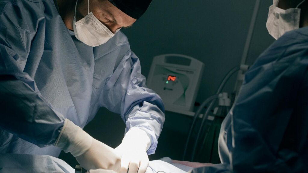
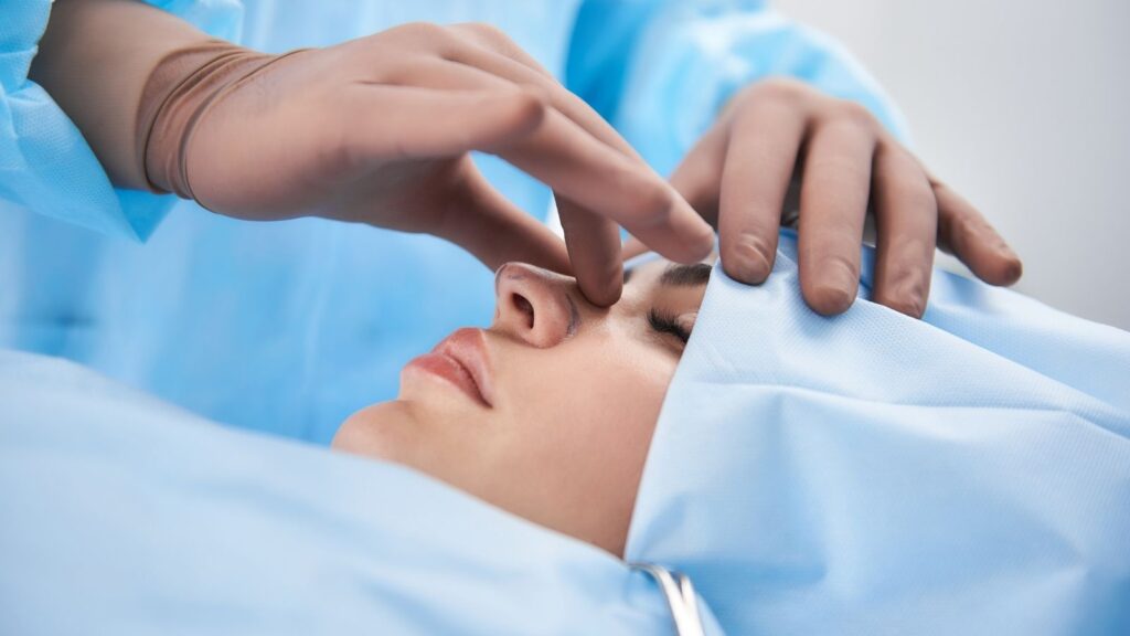
Nasal valve stenosis surgery is performed with techniques aimed at widening the intranasal airway. The valve area is usually opened using cartilage grafts or structural support materials. The operation is carefully planned to provide both functional improvement and aesthetic balance.
During the postoperative recovery period, swelling and mild discomfort may occur. Regular check-ups, proper nasal care, and adherence to the surgeon’s recommendations increase the success of the operation. With early diagnosis and proper treatment, patients can achieve healthy, comfortable, and functional breathing.
What Is Nasal Valve Stenosis?
Nasal valve stenosis is a breathing problem that occurs due to narrowing of the nasal valve area, which is the narrowest air passage inside the nose. This condition prevents sufficient airflow through the nose and causes a feeling of congestion especially while inhaling. It can generally develop due to congenital structural problems, trauma, or previous nasal surgeries. In treatment, the valve area can be widened using surgical methods.
Why is nasal valve stenosis e so critical for our breathing?
More than half (50–75%) of the resistance against the flow of air through our nose occurs in this small area called the “nasal valve region.” In other words, no matter how large or open your nose is, if there is a problem in this narrow area, you cannot breathe comfortably. This is not a single anatomical point but a three-dimensional, dynamic structure consisting of cartilage, bone, and soft tissues that work in harmony. To understand this region better, we can divide it into two main parts.
Internal Nasal Valve (İNV): It is the narrowest point of the nasal cavity. It is where airflow accelerates the most and pressure drops the most. The geometry of this area is vital for healthy breathing. Its boundaries are as follows:
- Medially, the nasal septum
- Superiorly and laterally, the inferior edge of the upper lateral cartilages
- Inferiorly and laterally, the head of the inferior turbinate (commonly known as “nasal concha”)
- Inferiorly, the nasal floor
External Nasal Valve (DNV): Refers to the area right at the entrance of the nostrils. Whether our nasal wings collapse inward during deep breathing gives us an idea about the health of this region. The main structures forming the structural integrity of this area are:
- Medially, the most anterior part of the nasal septum
- Laterally, the cartilages forming our nasal wings (lower lateral cartilages)
- Inferiorly, the nasal sill
A structural weakness or narrowing in either of these two regions can severely obstruct the airway. Therefore, successful treatment requires a comprehensive approach that evaluates these regions as a whole, rather than focusing on a single problem (for example, only a septal deviation).
How does nasal valve stenosis that causes my nose to collapse while breathing occur?
Behind the situation many of us experience—especially while exercising or taking a deep breath—where the nasal wings adhere inward and abruptly cut off the breath, lies a fundamental physical principle. When air passes rapidly through the nasal valve area, which is a narrow channel, it reduces the internal pressure in that region. Meanwhile, since the atmospheric pressure outside the nose remains constant, this “vacuum” effect pulls the nasal sidewalls inward if the cartilage support is weak. We call this “dynamic collapse.”
This principle causes nasal valve stenosis to present in two different forms. Making this distinction is very important to plan the correct treatment.
Static Nasal Valve Obstruction: In this case, the problem is a structural narrowing that is always present, independent of breathing. The airway is anatomically narrow even at rest. Some factors leading to this include:
- A septal deviation corresponding to the valve region
- Excessive enlargement of the head of the inferior turbinate
- Congenitally narrow bony nasal structure (piriform aperture)
- Scar tissue resulting from previous surgeries or trauma
Dynamic Nasal Valve Obstruction: In this case, the nasal structure may be sufficiently open at rest. However, when the person breathes in, the weak sidewalls, which cannot withstand the negative pressure, collapse inward. In other words, the problem is a “load-bearing system weakness” rather than a structural narrowing. Some causes leading to this include:
- Weak or malpositioned nasal wing cartilages
- Weakening of the flexible connections between cartilages (scroll area)
- Loss of the natural resilience of the cartilages with aging
Most of the time, these two conditions coexist in patients. That is, there is both an existing narrowing and an additional collapse that increases with breathing. Addressing both problems during surgical planning is essential for a lasting solution.
What are the main causes leading to nasal valve stenosis?
Nasal valve stenosis is generally an acquired condition and there are several underlying main causes. Knowing these causes is important both for prevention and accurate diagnosis. The main etiological factors are:
Previous Nasal Surgeries: Unfortunately, the most common cause of nasal valve problems is previous nasal surgeries, especially cosmetic (rhinoplasty) operations. Procedures focused solely on reduction and narrowing, without preserving the nose’s support mechanisms, can weaken the nasal framework. Maneuvers such as over-resection of the nasal hump, excessive removal of cartilages, or over-narrowing of the nasal bones can irreversibly weaken the valve area. This demonstrates how vital the principle “the primary goal of every aesthetic nasal surgery should be to preserve and improve function as much as aesthetics” is.
Trauma: Blunt impacts to the nose, sports injuries, or accidents can break, displace, or crush the delicate cartilage structures that form the valve region. This can cause an obstruction that appears immediately after the trauma or gradually as scar tissue matures over months.
Congenital and Structural Predisposition: Some individuals are more prone to nasal valve stenosis due to congenital anatomical features. For example, tight and narrow nostrils, an excessively projecting or drooping nasal tip, congenitally weak and thin cartilages, or a Caucasian-type narrow nasal structure can predispose a person to this problem.
Aging Process: Just like our skin, our cartilages lose their elasticity, hydration, and structural resilience with age. Cartilages that weaken and sag over the years can cause the nasal tip to drop and the valve region to gradually narrow under the effect of gravity. Therefore, even in people who have never had nasal problems before, slowly developing nasal obstruction may be seen in advanced age.
What symptoms might indicate that I have nasal valve stenosis?
The symptoms of nasal valve stenosis are quite typical, and the fact that a person experiences temporary relief with certain self-performed maneuvers provides an important clue for diagnosis. If you are experiencing one or more of the following symptoms, the source of your problem may be the nasal valve:
- Persistent nasal obstruction or obstruction that increases with exertion
- Nasal wings adhering inward especially while taking a deep breath
- While lying down, complete closure on the side that remains below, especially when lying on your side
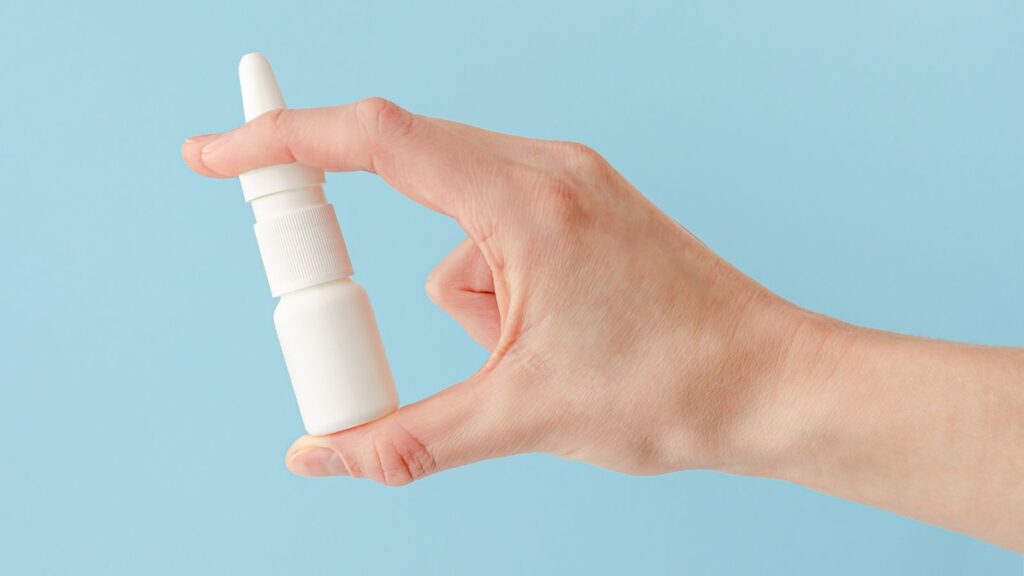
- Stubborn feeling of congestion that does not improve with nasal decongestant sprays
- Constant need to breathe through the mouth
- Dry mouth and throat irritation
- Snoring and deterioration in sleep quality
- Daytime fatigue and difficulty concentrating
- Significant opening of breathing when using over-the-counter nasal strips
- A thin whistling sound heard from the nose while breathing
In addition to these symptoms, a simple test called the “Cottle maneuver” can also guide you. With your index finger, gently pull your cheek outward from just beside your nasal wing. If you feel a marked opening and relief in your breathing with this simple movement, this is a strong sign of nasal valve stenosis.
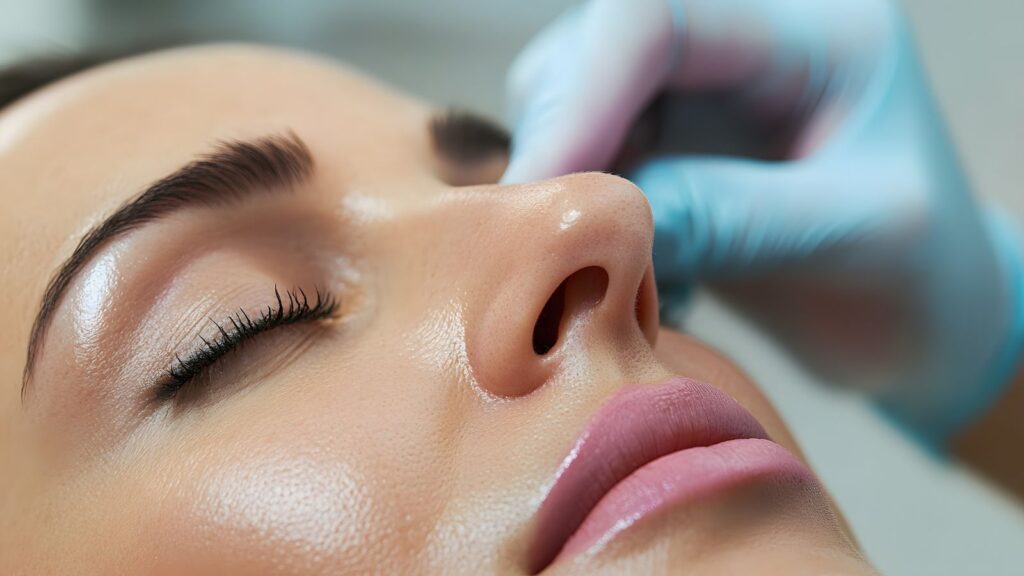
How does a doctor diagnose nasal valve stenosis and what tests are ordered?
The diagnosis of nasal valve stenosis relies more on the physician’s experience, careful examination, and accurate interpretation of the patient’s statements than on technological devices. It is essentially a clinical diagnosis. The steps of the diagnostic process are generally as follows:
- Detailed Patient History: Your doctor thoroughly inquires when and how your complaints started, under what conditions they increase, whether you have had previous nasal surgery or trauma, and how you responded to tests such as the Cottle maneuver.
- Physical Examination: This is the most important step. Your doctor carefully observes the external structure of your nose, asking you to take both normal and deep breaths. They evaluate whether there is any collapse in the nasal wings or sidewalls.
- Modified Cottle Maneuver: In addition to the standard maneuver, your doctor uses a small cotton-tipped applicator or blunt instrument to gently support first the external valve (nasal wing) and then the internal valve region from inside your nose, checking changes in your breathing. This is very helpful in determining from which point the collapse originates.
- Endoscopic Evaluation: Using a thin endoscope with a camera, the entire nasal cavity is examined in detail. The primary purpose of this examination is not to confirm the presence of collapse in the valve region, but to rule out other pathologies that may cause obstruction (such as posterior septal deviation, polyps, sinusitis, adenoid tissue, or tumors). It is essential to view the entire nose for a comprehensive treatment plan.
Are CT or MRI Necessary?
No. Radiological imaging methods such as computed tomography (CT) or magnetic resonance imaging (MRI) are not routinely required to diagnose dynamic valve collapse that occurs with breathing. These tests capture a snapshot of a static moment and cannot show cartilage movement and collapse during inhalation. Imaging methods are requested only to detail an additional bony or sinus pathology suspected during endoscopic examination.
What surgical techniques are used for internal nasal valve stenosis?
The main goal of surgical techniques applied to correct narrowing or collapse in the internal valve region is to permanently widen the narrowed air passage and/or provide support to the weakened cartilaginous framework. The method to be chosen is determined according to the severity of the problem and the anatomical structure.
- Spreader Grafts (Expanding Grafts): This technique is considered the “gold standard” of internal valve repair. Flat and sturdy strips, usually taken from the patient’s own nasal septal cartilage, are used. These cartilage strips are placed like a “support post” between the septum and the upper lateral cartilages on the nasal dorsum. The main objectives of this procedure are:
Pushing the upper lateral cartilages outward to widen the narrowed valve angle
Repairing an “open roof” deformity in patients whose nasal hump has been removed and preventing middle vault collapse
Acting as a splint to bring a crooked nasal dorsum into a straight line
- Butterfly Graft: This is a “scaffold” technique that is particularly effective in dynamic collapses. A butterfly-shaped piece is prepared from the patient’s ear cartilage, which is often preferred due to its natural curvature. This graft is placed under the skin, directly over the valve region where the collapse occurs. Working like an “internal nasal strip,” it prevents the cartilages from collapsing inward during breathing. This technique has advantages and disadvantages:
Advantages: It is very powerful in correcting dynamic collapse.
Disadvantages: There is a risk of creating fullness or a bulge on the nasal dorsum and just above the tip. Therefore, the graft must be finely adjusted.
Suture Techniques (Flaring Sutures): This is a method of reshaping cartilages using sutures alone, without grafts. By using special suture techniques, the upper lateral cartilages are pulled outward and the valve angle is widened “like a fan.” This method may be preferred especially in mild to moderate dynamic collapses and in situations where additional thickness in the nose is not desired.
How is surgery for external nasal valve stenosis and nasal wing collapse performed?
Problems in the external valve usually stem from weakness of the nasal wings or deformities of the nasal tip cartilages. The aim here is to provide permanent support to these collapsing structures.
Alar Batten Grafts: This is one of the most frequently used and most reliable methods in the treatment of external valve collapse. A piece of cartilage is placed on the weak lateral wall exactly at the point of maximum collapse, like a “support patch” or “beam.” By supporting the wall against the negative pressure that occurs during breathing, this graft prevents collapse. The main goals of this technique are:
- Strengthening the lateral wall experiencing dynamic collapse
- Filling the deep “cheek–nose groove” formed due to collapse to provide a smoother aesthetic contour
- Providing structural support to the nasal wing
Lateral Crural Strut Graft (LCSG): This is a more advanced technique and is used when there are not only weaknesses but also serious deformities such as inward bending, collapse, or malposition of the nasal wing cartilage. In this technique, a strong and straight piece of cartilage taken from the patient’s own septum or rib is placed under the problematic wing cartilage like an “I-beam.” This strong support both straightens and reinforces the weak cartilage, correcting both function and nasal tip aesthetics simultaneously.
Alar Rim Grafts: Sometimes the collapse occurs only in the soft tissue portion without cartilage, at the very bottom edge of the nostril. To correct this, very thin and small cartilage strips called “rim grafts” are placed into a narrow tunnel created under the skin at the nostril rim. The aims of this technique are:
- Stiffening the nostril rim to prevent its collapse
- Correcting the notched appearance called “alar retraction”
- Providing symmetry in the nostrils
Is there a non-surgical or simpler solution for nasal valve stenosis?
Yes, thanks to technology in recent years, minimally invasive methods—offering excellent alternatives to traditional surgeries for selected patient groups—have been developed. These methods are generally ideal for patients who do not have a major structural deformity or severe bony deviation, but whose main problem is dynamic collapse of the nasal sidewalls while breathing. These procedures can mostly be performed under local anesthesia in office or clinic settings. Some advantages of these modern approaches are:
- No need for general anesthesia
- Much faster recovery
- No need for materials like packing or splints
- Short procedure time
- Minimal pain and discomfort
- Two main minimally invasive methods stand out.
Temperature-Controlled Radiofrequency Treatment: This method is based on delivering low-temperature radiofrequency energy into the submucosal layer of the nasal valve region via a special handheld device. This controlled heat provides a kind of “stiffening and reshaping” without cutting or removing tissue. With this procedure, the collapsing weak cartilage and soft tissue become more resistant and stable. Scientific studies have shown that in appropriate patients, this method can be as effective as traditional surgery and that the benefit can persist for up to 4 years.
Resorbable Nasal Implants (Latera): In this technique, via a small intranasal incision, a resorbable implant is placed to support the collapsing lateral wall. This implant provides mechanical support for approximately 18–24 months. During this period, the body forms its own supportive scar tissue around the implant. Even after the implant fully resorbs, this newly formed fibrotic tissue continues to support the wall. Numerous scientific studies have proven that this method is safe and effective, providing lasting and significant improvements in patients’ breathing scores.
Thanks to these minimally invasive methods, our treatment options are now tiered like a staircase. A large, comprehensive surgery is no longer the only option for every patient. If the main problem is isolated dynamic collapse, starting with these simpler and less risky office procedures may be the most appropriate approach.
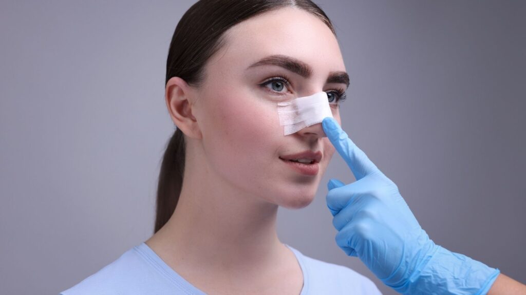
How does the recovery process progress after nasal valve surgery?
For a successful outcome, postoperative care is at least as important as the surgery itself. While the recovery process varies from person to person, there are some important points and restrictions that generally need attention:
Things to Do:
- Keep your head elevated with several pillows, even while sleeping, especially for the first few days.
- Clean your nose regularly with saline sprays or rinses as recommended by your doctor.
- Use the prescribed ointments as directed.
- When you need to sneeze, keep your mouth open to reduce pressure.
- Drink plenty of fluids.
Things to Avoid:
- Do not blow your nose at all for the first 1–2 weeks.
- Avoid strenuous activities such as sports, running, and weightlifting for at least 2–3 weeks.
- Do not lift anything heavier than 4.5 kg.
- Avoid bending forward or straining.
- Avoid swimming in pools or the sea for at least 4–6 weeks.
- Protect the surgical area from trauma.
- If you wear glasses, you may need to take a break to avoid pressure on the nasal bridge for a while.
At the end of the first week, the external cast on your nose and, if present, internal silicone splints are removed. However, this does not mean the healing is complete. Although much of the swelling subsides in a few weeks, it can take 6 months to 1 year, and sometimes even longer, for the nose to achieve its final shape and functional result. Being patient during this process and not missing your doctor’s follow-ups is the key to achieving the best outcome.
Frequently Asked Questions
What are the most common symptoms of nasal valve stenosis?
Nasal valve stenosis often causes persistent nasal obstruction, difficulty breathing through the nose, congestion, and reduced airflow. Symptoms may worsen during exercise, deep inhalation, or when lying down.
How can nasal valve stenosis affect daily activities and sleep quality?
Restricted airflow from nasal valve stenosis can contribute to mouth breathing, snoring, poor sleep quality, and daytime fatigue. Some patients also notice reduced exercise tolerance due to breathing difficulties.
What causes nasal valve stenosis to develop?
Nasal valve stenosis may result from congenital anatomy, previous nasal surgery, trauma, aging-related tissue weakening, or excessive scar formation. These factors can narrow the airway and restrict normal airflow.
How is nasal valve stenosis diagnosed by a specialist?
Diagnosis typically involves a physical examination, assessment of nasal airflow, and evaluation of nasal structures. Specialists may use endoscopic examination and functional tests to identify the location and severity of narrowing.
When is surgery recommended for nasal valve stenosis?
Surgery is generally considered when symptoms persist despite conservative treatments and significantly affect breathing. The decision depends on the severity of obstruction, anatomical findings, and overall patient health.
What surgical techniques are commonly used to correct nasal valve stenosis?
Procedures may include cartilage grafting, nasal valve reconstruction, structural support techniques, or functional rhinoplasty. The chosen approach depends on the cause of the narrowing and the patient’s anatomy.
Can nasal valve stenosis surgery improve both breathing and appearance?
In many cases, functional nasal surgery can enhance airflow while maintaining or improving nasal aesthetics. Treatment plans are often tailored to address both functional concerns and cosmetic goals when appropriate.
What is the recovery process like after nasal valve stenosis surgery?
Most patients experience temporary swelling, congestion, and mild discomfort during the early recovery period. Breathing improvements often become more noticeable as swelling decreases over the following weeks and months.
Are the results of nasal valve stenosis surgery permanent?
Results are generally long-lasting when structural support is successfully restored. However, healing patterns, tissue quality, aging, and individual anatomical factors can influence long-term outcomes.
What are the potential risks and complications of nasal valve stenosis surgery?
Possible risks include bleeding, infection, asymmetry, persistent obstruction, scarring, or dissatisfaction with the outcome. Careful surgical planning and postoperative follow-up help minimize these potential complications.