Rhinoplasty (nose aesthetic surgery) and dental treatments in Turkey, especially orthodontics and jaw surgery, cannot be considered separately in facial aesthetic planning. The fundamental relationship between these two fields comes from the fact that the upper jawbone (maxilla) forms the anatomical foundation on which the nose is seated. Any change in the position, width, or alignment of the teeth directly affects the posture, projection, and overall balance of the nose on the face. Therefore, a holistic aesthetic approach evaluates nasal and smile harmony together. Achieving the ideal result requires these two structures to be in complete harmony with each other.
What Is the Anatomical Connection Between the Nose and Jaw Structure?
To understand the relationship between the nose and jaw structure, we can make a simple analogy: Our upper jawbone (called the maxilla in medical terminology) is the foundation on which our nose is placed. Just as the stability and shape of a building depend on its foundation, the posture, shape, and support of the nose largely depend on the jawbone underneath.
This connection is not only theoretical but entirely physical. The floor of the nasal cavity—its lower base—is formed by the palate, which is part of the upper jawbone. A significant portion of the lateral walls of the nose also consists of the inner surfaces of this jawbone. In fact, the very bottom and front part of the “septum,” the cartilage-bone wall that starts between the eyebrows, extends to the nasal tip, and divides the nose into two, rests on a small bony projection in the upper jawbone called the “anterior nasal spine.” This projection plays a vital role in supporting and positioning the nasal tip. Therefore, any change in the position or size of the upper jaw directly affects the platform on which the nose sits.
This bony foundation forms a more complex network through muscles, vessels, and nerves. For example, some facial expression muscles that shape our smile, lift the upper lip, or move the nasal wings originate directly from the upper jawbone and attach to the nasal cartilages. This clearly shows how jaw position affects nasal tip mobility and appearance during smiling.
Clinically, one of the most important connections is the close relationship between the upper jaw sinuses and the posterior molar teeth. The bases of these air-filled cavities (sinuses) located inside the upper jaw are also where the roots of the upper molars lie. This distance is so small that, in most people, there is only a millimetric bone layer between the roots and the sinus floor. Sometimes the roots even extend directly into the sinus cavity. This close connection explains why a dental infection in this area can cause sinusitis, or why the sinus may be damaged during tooth extraction or implant placement. Therefore, this anatomical relationship must be carefully evaluated before any surgery involving this region.
How Do Breathing Habits and Dental Structure Affect Each Other?
The relationship between the nose and jaw is not only structural but also functional. Modern medicine considers the entire airway—from the nose to the sinuses and all the way to the lungs—as a “unified system.” Its main function is to clean, warm, and humidify the air before it reaches the lungs. Proper execution of this function largely depends on breathing through the nose.
So why is nasal breathing so important, and how is it related to dental structure? The answer lies in facial development during childhood. Imagine a child who cannot breathe comfortably through the nose due to adenoid hypertrophy, severe allergies, or a deviated septum and is forced to breathe through the mouth constantly. This is not merely a habit but triggers a chain reaction that negatively affects the development of the facial and jaw skeleton.
Continuous mouth breathing prevents the tongue from resting in its normal position on the palate, causing it to stay lower. The tongue, when resting on the palate, acts as a mold that helps the upper jaw develop laterally. Without this support, the upper jaw narrows, the palate deepens, and becomes high-arched. This reduces the space available for the teeth, leading to crowding. At the same time, a constantly open mouth and altered muscle balance cause the face to elongate vertically and the lower jaw to move backward. This condition is known as “adenoid face,” characterized by a long face, narrow upper jaw, and malocclusions (especially anterior open bite or posterior crossbite).
This relationship is bidirectional. A structurally narrow upper jaw can reduce the volume of the nasal cavity, leading to nasal obstruction and forcing the person into mouth breathing. This creates a vicious cycle: nasal obstruction disrupts jaw development, and altered jaw structure worsens nasal obstruction. Therefore, early diagnosis and treatment of nasal obstruction in children are crucial to preventing future complex orthodontic treatments or jaw surgeries. Similarly, in adults, orthognathic surgeries that expand or advance the upper jaw have been scientifically proven to significantly improve breathing by widening the nasal airway.
What Is the Importance of Nasal, Lip, and Jaw Proportions in Facial Harmony?
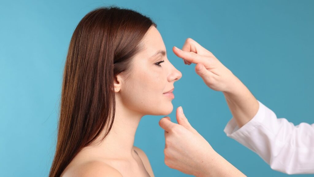
Facial beauty does not depend on the perfection of a single organ but on the harmonious proportions of all facial components. Because the nose is located at the center of the face, it is a key element of this balance. However, nasal aesthetics cannot be evaluated solely by the shape of the nose itself. Its relationship with the forehead, lips, and jaw determines how natural and harmonious the final result will be. These relationships are evaluated based on certain mathematically defined angles and proportions.
Some of the best-known ones include:
- Nasofrontal Angle: The angle at the junction of the forehead and nasal root. If it is too flat or too deep, it directly affects the profile view.
- Nasolabial Angle: The angle formed at the junction of the nasal tip (columella) and upper lip. It is one of the most important factors determining how upturned or droopy the nasal tip appears, and its ideal values differ for men and women. Women typically prefer a slightly wider (more upturned) angle, while men prefer a straighter, more masculine angle.
- Nasofacial Angle: The angle between the nasal dorsum and the vertical plane of the face. It expresses how projected or flat the nose appears.
These soft tissue angles and proportions reflect the underlying skeletal structure. For example, in a person with a retruded upper and lower jaw, the nose may appear larger and more projected (convex profile). Conversely, in a person with a more prominent jaw structure, the nose may appear flatter or even recessed (concave profile). This reveals a critical truth for surgical planning: simply reshaping or reducing the nose cannot always achieve the ideal result. Sometimes the real issue is jaw retrusion or protrusion, and unless this is corrected, facial imbalance may persist even after an excellent rhinoplasty. Therefore, a holistic approach that includes additional procedures such as chin augmentation (genioplasty) or jaw surgery (orthognathic surgery) when necessary provides the most harmonious and lasting results.
What Does the Preoperative Evaluation Process Include for a Successful Surgery?
Today, preoperative evaluation for rhinoplasty is much more comprehensive than simply taking photos and discussing what will be changed. This process includes factors ranging from patient expectations and psychological status to underlying health issues and dental/jaw structure, essentially creating a roadmap. The goal is to eliminate chance from the surgical outcome, foresee all possible risks, and ensure the patient is physically and psychologically ready for the process.
The first and most important step is honest and transparent communication between the patient and surgeon. The patient’s expectations, motivations, and reasons for wanting the surgery must be clearly understood. The surgeon’s role is to explain how much of these expectations can realistically be achieved surgically, what risks may exist, and what the recovery process truly entails. Unrealistic expectations are the leading cause of postoperative dissatisfaction, regardless of how technically successful the surgery is.
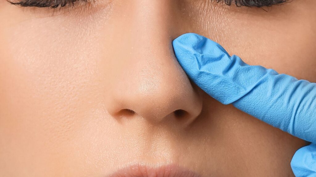
Another critical aspect is functional evaluation. The nose is not only an aesthetic organ but primarily a respiratory one. The patient must be evaluated for breathing problems, and a detailed intranasal examination should be performed. If there are issues such as septal deviation, turbinate hypertrophy, or nasal valve collapse, these should be addressed during the same surgery. Ignoring functional issues can worsen future breathing problems even after an aesthetically successful operation.
Which Health Conditions Should Be Screened Before Rhinoplasty?
For a safe surgical process and smooth recovery, every patient planned for rhinoplasty must be carefully screened for certain health conditions. Some important conditions that may affect the surgical plan include:
- Obstructive Sleep Apnea (OSA)
- Body Dysmorphic Disorder (BDD)
- Bleeding Disorders
- Chronic Nasal Spray Use
Sleep apnea is the repeated cessation of breathing during sleep and requires special precautions during anesthesia. Therefore, patients with complaints such as snoring or stopping breathing during sleep should be evaluated in this regard. Body dysmorphic disorder is a psychological condition in which a person becomes excessively preoccupied with a flaw that does not exist or is unnoticed by others. Since this condition may lead to dissatisfaction regardless of the surgical result, it must be considered in preoperative consultations. Conditions that increase bleeding tendency, such as blood-thinning medications or clotting disorders, must also be questioned, as they increase bleeding risk during and after surgery. Lastly, long-term and uncontrolled use of vasoconstrictor nasal sprays may cause permanent damage to the nasal mucosa and negatively affect the healing process. Therefore, such habits should be treated before surgery.
Why Is a Comprehensive Dental Examination Performed Before Surgery?
A comprehensive dental and periodontal examination is not optional but essential for patients who are planning rhinoplasty combined with jaw surgery. The primary reason is to minimize the risk of infection. The oral cavity contains billions of bacteria. Untreated tooth decay, an abscess in the tooth root, or advanced gum disease can serve as infection sources.
During major facial surgery, there is a risk of bacteria from these sources entering the bloodstream (bacteremia). These bacteria can reach the fresh surgical site and cause serious infection, loss of implants or plates placed during surgery, or even surgical failure.
To eliminate this risk, a detailed dental examination must be performed beforehand. In addition to clinical evaluation, this typically includes dental radiographs. If an active infection is detected, it must be treated before proceeding with major facial surgery. This requires close collaboration between the surgeon and the dentist and proper scheduling. This step is essential to ensure the patient’s overall health and the success of the surgery.
Which Modern Imaging Technologies Are Used in Surgical Planning?
One of the greatest revolutions in modern surgery is the ability to plan and simulate the procedure in a virtual environment even before the patient enters the operating room. This is particularly valuable in regions with complex three-dimensional anatomy such as the nose and jaw, increasing surgical precision and predictability. Some fundamental technologies used in this process include:
- Cone Beam Computed Tomography (CBCT)
- 3D Surface Imaging
- Digital Dental Scans
CBCT provides highly detailed 3D images of the facial and cranial bones with far lower radiation than standard CT scans. This allows millimetric evaluation of bone asymmetries, deformities, airway width, and implant placement areas. 3D surface imaging systems (such as Vectra) create a digital copy—an “avatar”—of the patient’s face using special cameras without radiation. Possible results of planned rhinoplasty or jaw surgery can be simulated on this model, helping both the patient and surgeon align on realistic expectations and shared aesthetic goals. Digital dental scans create a flawless 3D model of the teeth and gums using intraoral laser scanners. When combined with CBCT data, they enable the creation of personalized surgical guides for jaw surgeries, increasing surgical precision and success. Together, these technologies create a “digital twin” of the patient, allowing the surgeon to plan and rehearse the surgery in detail.
How Should the Timing of Rhinoplasty and Jaw Surgery Be Planned?
For a patient requiring both rhinoplasty and jaw surgery, one of the most critical questions is: Should these surgeries be performed together or separately? The decision must balance patient comfort and optimal aesthetic and functional outcomes.
Performing both surgeries together has several advantages for the patient: a single anesthesia session, a single recovery period, and overall lower cost. However, the situation is more complex from a surgical standpoint. Upper jaw surgery (Le Fort I osteotomy) changes the foundation on which the nose sits and causes significant facial swelling (edema). This swelling can complicate the delicate adjustments and symmetry checks required during rhinoplasty and reduce the predictability of the result.
Therefore, the decision is based on specific surgical criteria. The goal is to align patient preference with the best functional and aesthetic outcome.
When Is It Appropriate to Combine Jaw and Nose Surgeries?
Performing both surgeries in the same session is preferred when rhinoplasty results can remain predictable. These situations usually include:
- Minimal upper jaw movement
- Surgery involving only the lower jaw
- Correction of changes caused by jaw surgery
- Repair of existing nasal deformities
The most important evidence-based criterion is the amount of upper jaw movement. If the upper jaw is advanced less than 4–5 mm or moved upward less than 2 mm, this minimal movement is not expected to significantly alter the nasal foundation or cause excessive swelling. In such cases, rhinoplasty can be safely performed during the same session. If the jaw surgery only affects the lower jaw, combining it with rhinoplasty is no problem since it does not affect the nasal base. In some cases, rhinoplasty is performed to correct undesired aesthetic changes caused by jaw surgery—for example, expansion of the nasal wings after upper jaw advancement.
When Should the Surgeries Be Performed Separately?
If jaw surgery has a high potential to compromise rhinoplasty results, performing the surgeries in two separate stages is the safest and most correct approach. Key situations requiring this include:
- Significant upper jaw movement
- Waiting for soft tissue healing
Large skeletal movements—such as advancing the upper jaw more than 4–5 mm or elevating it more than 2 mm—significantly alter the nasal foundation and cause prolonged swelling. Under such conditions, it becomes nearly impossible to perform a precise rhinoplasty or achieve symmetrical, natural results. In these cases, jaw surgery is performed first, followed by a healing period to allow swelling to resolve and soft tissues to adapt to the new bone structure. Scientific studies suggest this waiting period should be approximately 7 months (about 208 days). After this period, rhinoplasty can be performed on a stable and predictable foundation, providing much better results. This approach prioritizes aesthetic excellence over patient convenience.
Which Special Surgical Techniques Are Used in Combined Surgeries?
When rhinoplasty and jaw surgery are combined, the surgeon manages the challenges while also benefiting from unique opportunities. These surgeries are not merely two consecutive procedures but integrated maneuvers that complement each other. Some special techniques include:
- Access to the nasal base via Le Fort I osteotomy
- Changing the position of the anesthesia tube
- Alar cinch sutures to control nasal flare
- Lip and gum aesthetics (V-Y advancement technique)
- Reshaping the septum
During upper jaw surgery (Le Fort I), the jawbone is sectioned and moved downward, providing excellent visibility and access to the nasal base, the lower septum, and the nasal cavity entrance—areas normally difficult to reach. This allows corrections such as septal deviation or harvesting cartilage grafts without additional incisions. One technical challenge in combined surgeries is managing the anesthesia tube. Ideally, jaw surgery is performed with nasal intubation, while rhinoplasty requires oral intubation. To resolve this, the nasal tube is switched to an oral tube at the end of jaw surgery in coordination with the anesthesia team, a process completed within seconds by an experienced team. Upper jaw movement tends to widen the nasal wings, so alar cinch sutures are placed to control this widening. For patients with gummy smiles (excessive gum display while smiling), the V-Y advancement technique is used during closure to lengthen the upper lip. When the upper jaw is moved upward, the lower edge of the septum must be shortened to prevent buckling and breathing problems.
How Is Orthodontic Treatment Timed with Rhinoplasty?
For patients undergoing or planning orthodontic treatment, the timing of rhinoplasty is important. If braces are used only to correct minor crowding and do not significantly affect the facial profile, rhinoplasty can be performed while the braces are still in place.
However, if the goal of orthodontic treatment is to significantly change the position of the upper or lower jaw—for example, correcting a major malocclusion—this will alter the profile and soft tissue balance. In such cases, performing rhinoplasty before the new skeletal balance is established is like painting on a canvas whose final shape is unknown. Therefore, when orthodontic treatment will significantly affect the profile, it is best to wait until the treatment is completed and the facial structure stabilizes before planning rhinoplasty.
If rhinoplasty is performed first, orthodontic treatment (such as braces) should typically wait 6–8 weeks for initial swelling to subside and tissues to heal.
How Does Having a Dental Implant in the Upper Jaw Affect Rhinoplasty Planning?
In a patient with dental implants in the upper jaw—especially in the anterior region—rhinoplasty planning must consider the location of these implants. The apex of the implants may sometimes be very close to the nasal floor or even extend toward the nasal base. Rhinoplasty itself generally does not harm stable, osseointegrated implants. However, if any intervention is planned in the nasal base or anterior maxilla during surgery (for example, while placing support grafts for the nasal tip), the exact position of the implant must be known. Therefore, a preoperative CBCT scan is important to map the relationship between implants and nasal structures.
Conversely, if a patient planning rhinoplasty also needs a “sinus lift” due to insufficient bone for implant placement, these two surgeries should be appropriately timed. A sinus lift can cause temporary swelling and reactions in the nasal and sinus tissues. Therefore, after such major dental procedures, a waiting period of 4 to 9 months is recommended to allow tissues to heal completely before performing rhinoplasty.
Frequently Asked Questions
Can I Have Dental Treatment After Rhinoplasty?
Yes, dental treatment is possible after rhinoplasty, but it is generally recommended to wait until the initial healing period is complete. Your surgeon can advise you on the appropriate timing based on your recovery.
How Long Should I Wait Before Visiting the Dentist After Rhinoplasty?
Many surgeons recommend waiting at least four to six weeks before undergoing routine dental procedures, especially those that require significant mouth opening or pressure around the nose and upper jaw.
Can Dental Work Affect Rhinoplasty Results?
Certain dental procedures may place pressure on facial structures and potentially cause discomfort during the healing process. Following your surgeon’s recommendations can help protect your surgical results.
Is It Safe to Have a Dental Cleaning After Rhinoplasty?
Routine dental cleanings are generally safe once your surgeon confirms that sufficient healing has occurred. It is important to inform your dentist about your recent surgery.
What Should I Tell My Dentist Before Treatment?
You should inform your dentist that you recently underwent rhinoplasty, provide details about your recovery, and discuss any restrictions recommended by your surgeon.
Can Orthodontic Treatment Be Continued After Rhinoplasty?
In most cases, orthodontic treatment can continue after rhinoplasty, but coordination between your orthodontist and surgeon may be beneficial to ensure a smooth recovery.
Will Dental Procedures Cause Pain After Rhinoplasty?
Some patients may experience temporary discomfort if dental treatment is performed too soon after surgery. Waiting until healing progresses can help minimize sensitivity and irritation.
Can I Undergo Major Dental Surgery After Rhinoplasty?
Major dental procedures may require additional planning and timing considerations. It is best to consult both your surgeon and dentist before scheduling extensive dental work.
Why Is Timing Important Between Rhinoplasty and Dental Treatment?
Proper timing allows the nasal tissues and surrounding structures to heal without unnecessary pressure, reducing the risk of discomfort or complications during recovery.
Should My Surgeon and Dentist Communicate About My Treatment?
Yes, communication between your surgeon and dentist can help ensure that both treatments are coordinated effectively and that your recovery remains safe and successful.