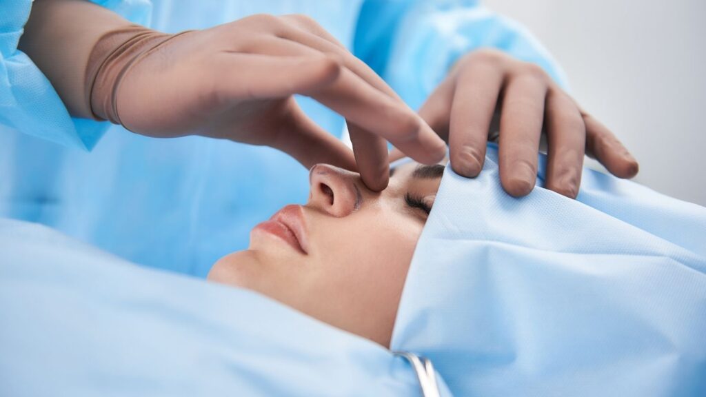
Rhinoplasty, commonly known as nose aesthetic surgery, is a facial aesthetic surgery procedure that aims to improve both the cosmetic appearance of the nose and its functional features such as breathing. Ear aesthetics, or otoplasty, on the other hand, most often corrects the “protruding ear” deformity by reshaping the ears’ form, size, and angle relative to the head, thus enhancing the overall harmony and symmetry of the face. Both operations aim to achieve natural and balanced results that are compatible with the person’s facial features, thereby addressing aesthetic concerns and positively impacting self-confidence.
Is rhinoplasty only cosmetic, or does it also address breathing problems?
This is one of the most fundamental questions in the minds of almost everyone considering a rhinoplasty journey. The answer, in the context of modern surgical understanding, is very clear: aesthetics and function—that is, an attractive appearance and healthy breathing—are a whole that can never be considered separately. Imagine you have a stunning sports car, but its engine does not run. No matter how beautiful it looks, it falls short because it cannot fulfill its primary function. The nose is just like that.
In the past, these two issues used to be handled separately. Surgeries performed for aesthetic concerns and those for improving breathing were different. However, we now know that the bones and cartilages that form the nasal framework also make up the walls of the internal air passages. These two structures are like the supporting columns and exterior façade of the same building. When you intervene in one, you inevitably affect the other.
For example, lowering the nasal hump too much for purely aesthetic reasons may narrow the critical area we call the “internal nasal valve,” which allows air to reach the lungs comfortably. The result? A nose that looks better but cannot breathe. Conversely, a cartilaginous deviation inside the nose (septal deviation) not only causes snoring or constant nasal congestion, but can also make the nose appear crooked, twisted, or asymmetrical from the outside.
For this reason, a successful rhinoplasty today stands on three pillars.
The key elements that determine patient satisfaction are:
- Functional satisfaction (being able to breathe comfortably)
- Cosmetic satisfaction (a natural appearance in harmony with the face)
- Psychological satisfaction (increased self-confidence and social ease)
Even a technically flawless operation cannot be considered truly successful if it does not align with the patient’s expectations or creates a new functional problem. Therefore, rhinoplasty is not merely the reshaping of an organ, but the art of improving overall quality of life.
Why does the pre-rhinoplasty consultation play such a critical role?
The initial preoperative consultation is the most important step in the entire process. It is not just a meeting, but a planning session where the roadmap of the journey is drawn, trust is built, and dreams meet reality. A successful outcome is directly linked to the quality of this consultation.
During this visit, your surgeon aims to understand you and your nose in a holistic way. There are several indispensable steps in this process:
The main evaluation steps during the consultation are:
- Detailed medical history
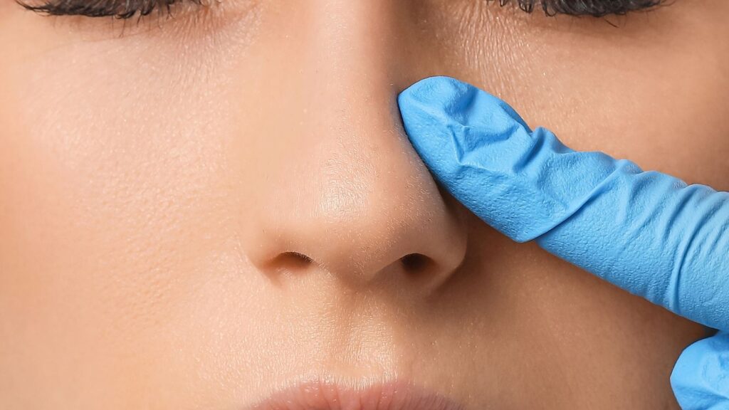
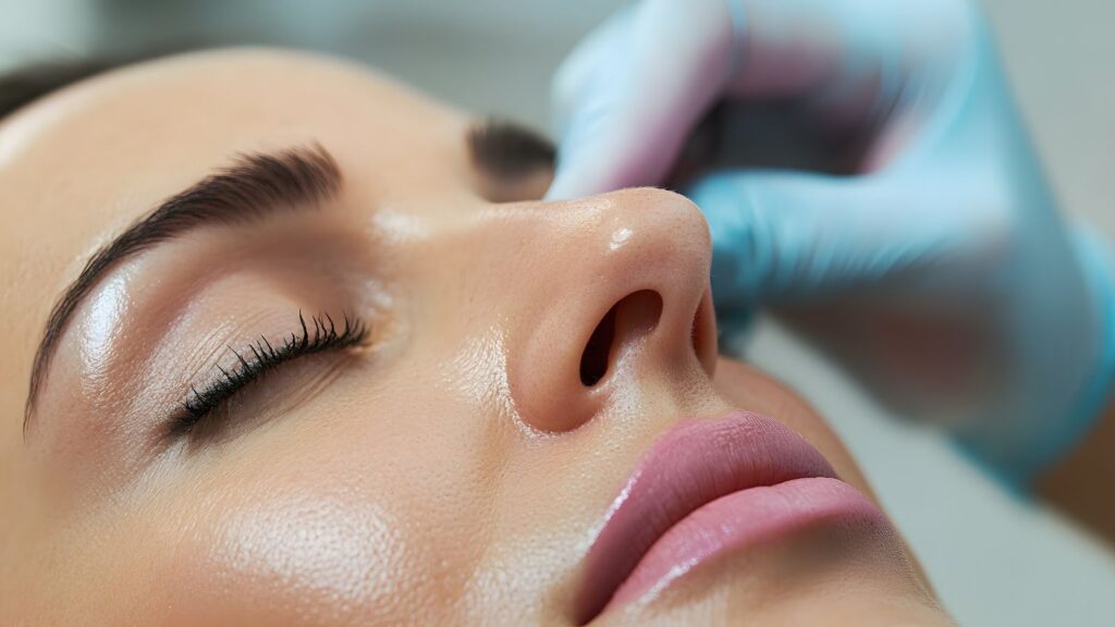
- Comprehensive physical examination
- Systematic facial and nasal analysis
- Professional photography
- Digital simulation and goal setting
First, your medical history is reviewed. Details such as previous surgeries, allergies, medications you use, and bleeding problems are vital for the safety of the operation. Then comes the physical examination. The inside of the nose is examined in detail with an endoscopic camera to look for answers to questions such as: Is there a deviation in the septum, what is the condition of the turbinates, are the sinus pathways open?
In the external examination, the anatomical structure of your nose is evaluated almost like a work of art. Imagine the skeletal framework of your nose as a piece of furniture and your skin as the cover over it. A thick, high-quality velvet cover hides the small imperfections underneath, while a thin silk cover reveals even the slightest scratch. In the same way, the thickness and quality of your nasal skin and the strength of the underlying cartilage matter. The surgeon must foresee how this “cover” will adapt to the new framework.
Afterward, photographs are taken from different angles and analyzed digitally. Today, 3D simulation technology has become an indispensable part of this stage. Thanks to this technology, you can see the potential results on your own face in three dimensions. You can visually explore answers to questions such as “How would I look if my nasal tip were slightly more elevated?” or “What will my profile look like if my hump is straightened?”. This allows you to express what you want to your surgeon in the clearest way, and enables your surgeon to show you what is realistically possible. Remember, this is not a guarantee but a shared target. Defining this shared target is the most important beginning of the journey.
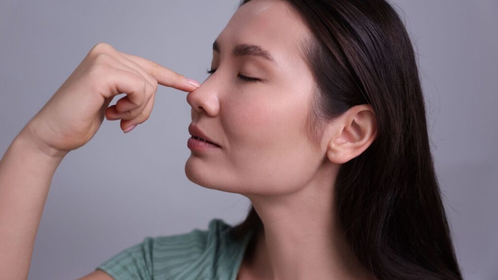
Why is the chin so important in facial aesthetics and rhinoplasty planning?
Evaluating the nose as an isolated structure, independent from the face, is like trying to understand a landscape by only looking at a single tree. What makes that tree meaningful is the mountain behind it, the river beside it, and the sky above it. Similarly, the nose gains meaning and aesthetic balance through its relationship with the other facial features, especially the chin. This holistic approach is called “profiloplasty,” or profile aesthetics.
In profile aesthetics, the most critical balance is the imaginary line between the nasal tip and the tip of the chin. Especially in the side profile, a small or retruded chin (microgenia) creates an optical illusion that makes the nose appear much larger, longer, and more projected than it really is. In such cases, although the patient may complain that “my nose is too big,” the real problem is the imbalance between the nose and the chin. A surgery that only reduces the nose may be technically successful, but still fail to correct the facial disproportions. The nose becomes smaller, yet the profile may remain weak and unbalanced.
In such situations, to achieve the most harmonious and aesthetic result, chin augmentation (genioplasty) can be considered alongside rhinoplasty. This procedure is usually performed by placing a biocompatible implant at the chin tip or by advancing the patient’s own chin bone. The goal is to give the lower face stronger definition and to balance the profile. Performing both procedures together can create a more dramatic and harmonious transformation than rhinoplasty alone. This requires the surgeon not only to operate on the nose, but also to read the entire face as a whole and to possess artistic vision.
What is the difference between open and closed rhinoplasty techniques?
This is also one of the topics patients are most curious about: “Which technique is better?” The most honest answer to this question is: “There is no universally better technique, there is only the technique that is more appropriate for your nose.” Open and closed techniques are not rivals; they are different paths the surgeon may use to reach the desired goal. Just as a carpenter sometimes uses nails and sometimes screws, the surgeon chooses the most appropriate path depending on the needs of the nose.
Closed (Endonasal) Rhinoplasty: In this technique, all incisions are made inside the nostrils. There is no visible scar on the outside. The surgeon works with a tunnel vision, that is, with a more limited field of view.
Advantages: Its greatest advantage is the absence of any external scar. Since the tissues are generally less traumatized, postoperative swelling and bruising may be slightly less, and recovery may feel somewhat faster.
Disadvantages: Because the field of view is limited, it is more difficult to place delicate sutures or cartilage grafts in complex cases. For this reason, it is usually preferred for noses that require simpler, smaller adjustments.
Open (External) Rhinoplasty: In this technique, in addition to incisions inside the nose, a small, usually “V”-shaped or step-shaped incision is made on the strip of skin that separates the nostrils, called the columella. Through this incision, the nasal skin is lifted like the hood of a car.
Advantages: It allows the surgeon to see the entire bony and cartilaginous framework directly and in great detail. This offers an unmatched level of control and precision. It is the gold standard for noses with significant deviation, those that have been operated on before (revision), asymmetric noses, or noses that need to be structurally rebuilt using cartilage grafts.
Disadvantages: A small scar remains on the columella. However, when the incision is properly closed using the right technique, this scar becomes almost invisible within a few months and is very difficult to notice. Swelling may be somewhat more pronounced compared with the closed technique.
At the point modern surgery has reached, this debate has largely lost its meaning. Large scientific studies have shown that both techniques can yield excellent results when applied to the right patients. What truly matters is that the surgeon is proficient in both methods and has the wisdom to choose the one that best suits your anatomical structure and aesthetic goals.
What does the philosophy of preservation rhinoplasty mean?
Preservation rhinoplasty is a modern philosophy that has become increasingly prominent in recent years, bringing a gentler, anatomy-respecting perspective to surgery. In contrast to the traditional “break-and-rebuild” (reductive) approach, preservation rhinoplasty is based on the principle of “reduce-and-reshape” (preservation).
Its core logic is this: Instead of disrupting the nose’s natural structures—especially the smooth, strong anatomical integrity of the nasal dorsum—it aims to reshape the nose while preserving them.
In the traditional method, the hump on the nasal dorsum is removed with a rasp or chisel. This creates what we call an “open roof” deformity, and the lateral nasal bones must then be fractured to close this roof. In the preservation technique, however, the dorsum itself is not touched. Similar to shortening a table by cutting its legs instead of sanding down the tabletop, small, millimetric segments are removed from the supporting structures beneath the dorsum (the septum and lateral walls). In this way, the entire nasal dorsum is lowered as a single block without disrupting its natural architecture.
There are several key principles at the heart of the preservation philosophy:
The main principles of this approach are:
- Preserving the integrity of the nasal dorsum
- Preserving the nasal ligaments
- Minimizing cartilage resection (removal)
This approach can yield excellent results particularly in patients with a pronounced dorsal hump but without major problems in the nasal tip or overall structure. It offers less trauma, less swelling and bruising, faster recovery, and most importantly, a nasal dorsum that looks extremely natural and “non-operated.” However, this technique is not suitable for every nasal anatomy and requires a significant learning curve. It is critically important for the surgeon to accurately determine which patients will benefit from this technique.
Why are revision rhinoplasty operations more demanding?
Revision rhinoplasty is a corrective operation performed on individuals who have undergone one or more previous nasal surgeries but have not achieved the desired result. These surgeries are among the most challenging and experience-dependent procedures in aesthetic surgery. There are several fundamental reasons for this:
There are key factors that make revision surgery difficult.
- Distorted anatomy
- Scar tissue
- Cartilage deficiency
- Reduced skin quality
- Psychological factors
In the first surgery, the natural anatomical planes and structures have been altered. The surgeon is no longer operating on untouched terrain, but on an area that has previously been modified, where the “roads” have changed and unexpected issues may arise. The dense, adherent scar tissue formed during the healing process makes it considerably harder to separate normal tissues from one another.
Perhaps the greatest difficulty is the lack of “building material.” Cartilage is required to reshape and support the nose. In the first surgery, the septum—the ideal source of cartilage in the middle of the nose—is often already used or damaged. In that case, the surgeon must find a new source. The second option is usually the ear cartilage. Cartilage taken from behind the ear can be used for small to medium-scale repairs. If greater and stronger support is needed, cartilage from the patient’s own rib may be required. This means an additional surgical incision and a separate healing process.
Besides all these technical challenges, the patient’s psychological state is also very important. The disappointment, anxiety, and distrust experienced after the first surgery make the process more delicate. Therefore, in revision rhinoplasty, the surgeon’s empathy, open communication, and ability to manage expectations realistically are at least as important as technical skill.
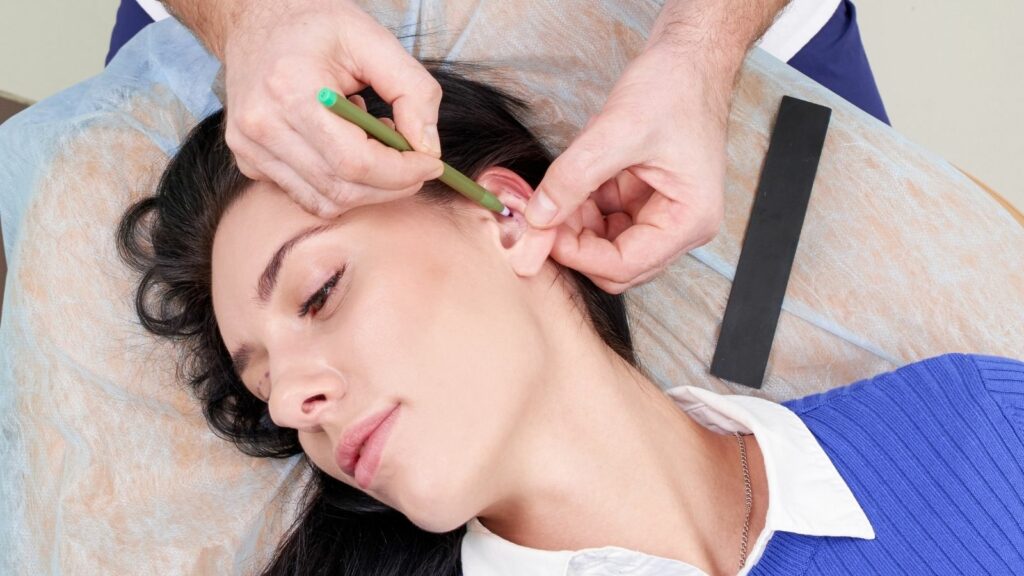
What are the main goals and reasons for protruding ear (otoplasty) surgery?
Otoplasty is a surgical procedure used to correct the condition commonly known as “protruding ears,” where the ears stick out from the head at a more pronounced angle than normal. Although this is not a medical health problem, it can deeply affect a person’s social life and psychology, especially during childhood and adolescence. Being teased, feeling the need to hide the ears with hair, and similar situations can lead to lack of self-confidence. The main goal of otoplasty is to give the ears a more natural, aesthetic, and facially harmonious appearance, thereby eliminating this psychosocial pressure.
There are two main anatomical causes underlying the protruding ear appearance:
The main causes of this deformity are:
- Insufficient antihelical fold: The “Y”-shaped natural fold that should be present in the upper part of the ear either does not form or is poorly defined. When this fold is absent, the ear appears flat and sticks outwards.
- Enlarged conchal cartilage: The bowl-shaped (concha) cartilage in the middle part of the ear is deeper or larger than normal. This pushes the entire ear forward away from the skull.
Most of the time, these two causes coexist. Surgical planning begins with correctly diagnosing which of these problems—or to what degree both—are present. The goal of surgery is not merely to stitch the ear back but to reshape these underdeveloped or overdeveloped anatomical structures in order to achieve a stable and natural result. The operation can generally be performed in children from around 5–6 years of age, when ear development is largely complete. Performing the surgery at this age can help the child overcome the issue before starting school and prevent potential psychological trauma.
What modern and effective techniques are used in otoplasty?
In protruding ear surgery, there are essentially two different philosophies: techniques that shape the cartilage by cutting or weakening it, and techniques that preserve the integrity of the cartilage and shape it only with sutures. Modern surgery is evolving day by day toward less traumatic methods that preserve the cartilage as much as possible.
In the past, techniques that involved scraping (scoring) the front surface of the cartilage or removing pieces of cartilage to reshape the ear were popular. Although effective, these methods carried the risk of creating sharp, unnatural edges or irregularities in the cartilage over time.
Today, the gold standard is suture-based techniques that preserve the cartilage. In this method, the entire procedure is performed through an incision made behind the ear. The cartilage itself is not cut. Instead, permanent, non-absorbable sutures are placed through the cartilage in strategic positions. When these sutures are tightened, the missing antihelical fold is naturally created and/or the enlarged conchal cartilage is repositioned closer to the head.
The main advantages of this technique are:
- The main benefits of the suture-based technique
- Extremely natural and smooth ear contours
- Preservation of cartilage integrity
- Easier revision surgery if needed in the future
In the most advanced version of this technique, the permanent sutures are covered with a thin layer of fascia taken from behind the ear. This almost completely eliminates the risk of the sutures being felt under the skin or eroding through it over time, and increases the longevity of the result. This “reinforced preservation” approach represents the modern understanding of otoplasty, combining the lowest complication rates with the most aesthetic outcomes.
What should be considered during the recovery period after rhinoplasty and otoplasty?
No matter how successful the surgery is, one of the most important factors determining the final outcome is the proper management of the postoperative recovery process. Each of these operations has its own unique healing rhythm:
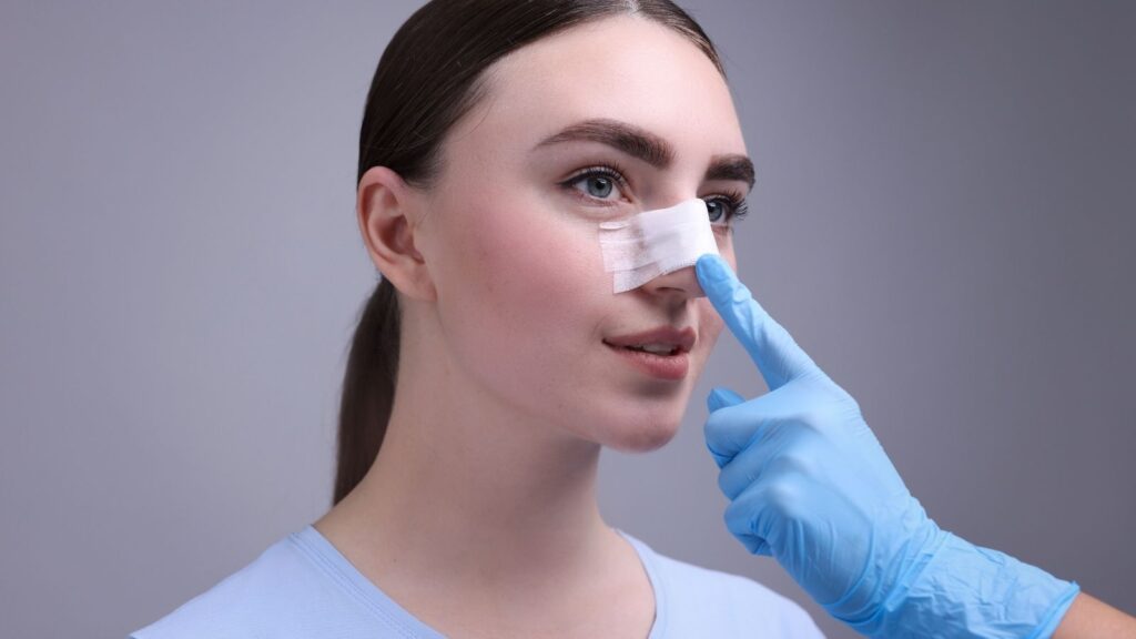
After Rhinoplasty: This process requires patience.
- First Week: A protective splint is placed on the nose. Swelling and bruising are at their peak. It is recommended to keep the head elevated and apply cold compresses.
- First Month: After the splint is removed, most of the swelling subsides quickly. However, the nose is still edematous and sensitive. It is very important to avoid heavy exercise, wearing glasses, and any trauma to the nose.
- First Year: The nose continues to refine and settle slowly until it reaches its final shape. Complete resolution of nasal tip edema can take up to a year. During this period, it is important to be patient and to know that the result will continue to improve day by day.
After Otoplasty: Recovery is usually faster and more comfortable.
- First Week: A bandage is used to wrap and support the ears in their new position. After this bandage is removed, a headband (sports/tennis band) is usually recommended for several more weeks, especially at night while sleeping, to protect the ears.
- First Month: Most of the swelling and bruising resolves. Patients can return to their normal daily routines. However, contact sports or activities that may cause pulling or trauma to the ears should be avoided for at least 4–6 weeks.
- After both operations, strictly following your surgeon’s instructions, not missing follow-up appointments, and remembering that healing is a marathon, not a sprint, are the keys to achieving the best possible result.