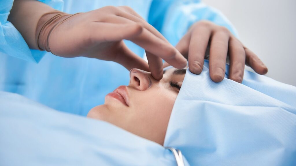
Intranasal adhesion surgery is the procedure of permanently opening air corridors blocked as a result of the opposing inner surfaces in the nasal cavity fusing together with abnormal scar tissue, using advanced technological methods. This surgical intervention aims to eliminate fibrotic bridges that completely block nasal physiology and to reconstruct the natural aerodynamics of the nose. This micro-reconstruction procedure, which not only eliminates a mechanical narrowing, directly improves quality of life by restoring healthy breathing function. Thanks to a successful operation, the blocked respiratory passages are freed, and the nose’s most basic functions of filtering, warming, and humidifying the air return completely to normal.
Why Does Intranasal Adhesion Occur and How Does the Need for Intranasal Adhesion Surgery Arise?
The process of intranasal adhesion formation is a highly complex pathological process that begins with the disruption of the physical integrity of the inner lining of the nose, which we call the mucosa, and unfortunately deviates from the natural wound-healing cycle. The nasal mucosa is one of the tissues in the human body with the highest capacity for self-renewal. However, when this tissue undergoes a surgical intervention or receives external physical trauma, the healing mechanism may not always proceed in the desired direction. This unwanted condition is quite likely to be encountered especially after bone and cartilage surgeries performed to correct deviation of the nasal septum, surgical procedures aimed at reducing the turbinates, or endoscopic interventions applied for the treatment of chronic sinusitis.
After any surgical trauma, a natural inflammatory process begins inside the nose. During this critical period, vascular permeability between cells increases, and a rich fluid that will initiate the repair process is released into the tissue space. The protein called fibrinogen, which is found abundantly in this repair fluid, forms fibrin networks on wound surfaces. These networks are actually microscopic temporary healing bridges. In a normal and healthy healing process, the rhythmic movements of the ciliated cells inside the nose and the natural secretion flow of the nose gently clear these temporary bridges and allow the tissues to heal independently.
However, if two opposing wounded surfaces are very close to each other and the cleaning mechanism between them is disrupted, a major problem begins. Factors such as intense tissue edema after surgery, supportive materials placed inside the nose remaining there for a long time, or the patient neglecting the nasal irrigations that must be performed after surgery directly impair this mechanism. These fibrin bridges, which cannot be cleared, begin to become permanent over time. In the following days, fibroblast cells, which are primarily responsible for tissue repair in the body, rapidly migrate to this area and begin intensive collagen production. Thus, those thin bridges that initially appear extremely soft and harmless harden over time and turn into cartilage-like fibrotic bands. From this point on, it becomes impossible for the tissues to open on their own, and surgical intervention becomes an essential need for the restoration of the anatomical structure.
How Does Intranasal Adhesion Impair Breathing and What Complaints Are Seen Before Intranasal Adhesion Surgery?
The negative effect of these fibrotic adhesions inside the nose on breathing cannot be evaluated only as a mechanical narrowing of space. This condition completely disrupts the entire aerodynamic structure of the nose, which has evolved over years. To understand how this mechanism is impaired, it is quite enlightening to look at the basic rules of fluid dynamics. Even millimetric narrowings occurring in the airways inside the nose suddenly and uncontrollably increase the flow rate of inhaled air, just like the pressure of water passing through a narrow pipe increases. At these narrow points where the speed increases abnormally, local air pressure suddenly drops. This pressure drop causes structurally more flexible and weaker areas, especially the nasal wings and nasal valve, to collapse inward with every breath.
Moreover, these uneven and narrow passages created by the adhesion completely prevent air from flowing smoothly and silently inside the nose. The airflow hits the adhesion areas, creating turbulence and vortices. This turbulent air continuously and very harshly strikes only certain areas of the nasal mucosa, causing that area to dry rapidly. The dried and cracked mucosa can no longer perform its protective function. The ciliated cells on it dry out and shed, and dense, uncomfortable crusting begins inside the nose. These dense secretions and hard crusts that cannot be cleaned create an extremely ideal, moist, and dark breeding environment for bacteria and fungi. This situation puts the patient into a difficult-to-break vicious cycle of constantly recurring, medication-resistant chronic sinusitis attacks.
As a reflection of these pathological processes, the problems patients encounter in their daily lives are quite evident. Depending on the size and location of the adhesion, the severity of symptoms may vary from person to person.
Commonly encountered complaints are as follows:
- Nasal congestion
- Mouth breathing
- Snoring
- Sleep apnea
- Morning fatigue
- Dry mouth
- Bad breath
- Headache
- Loss of smell
- Feeling of pressure in the face
What Are the Stages of Intranasal Adhesion and How Is the Decision for Intranasal Adhesion Surgery Made?
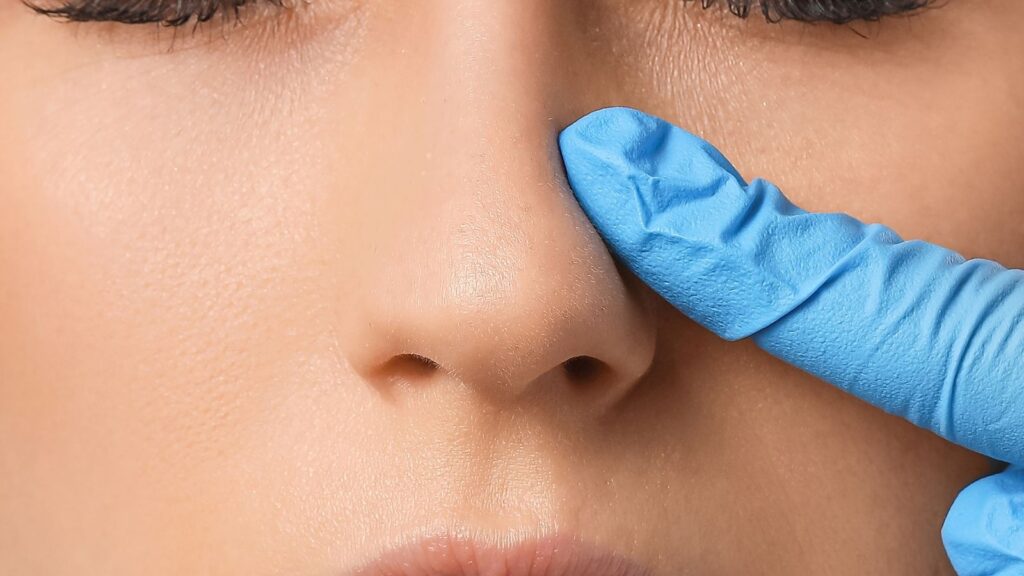
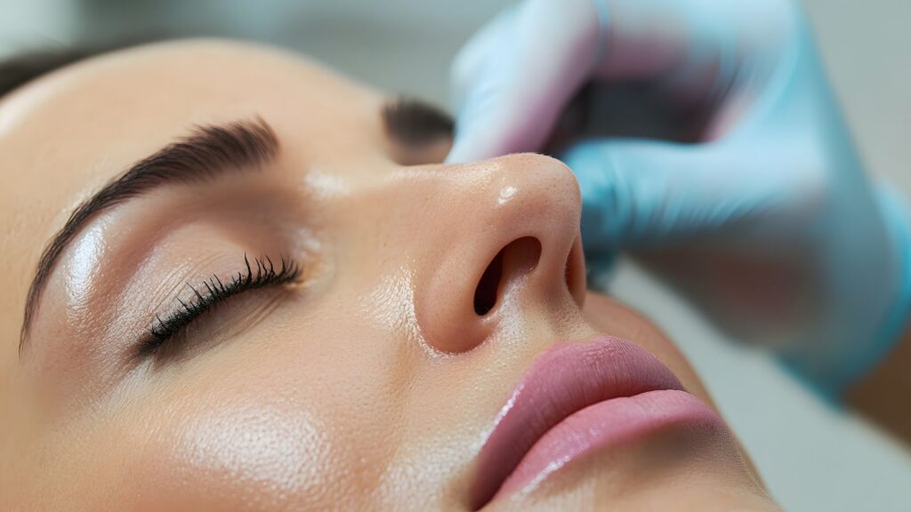
The management of intranasal adhesions in modern medicine begins with extremely accurate mapping of where the problem anatomically starts, where it extends, and how much volume it occupies. It is absolutely not possible to understand the size of the adhesion inside by simply looking at the nose from the outside. The gold standard of this detailed evaluation is high-resolution nasal endoscopy. Thanks to these thin, rod-shaped optical devices with a very powerful cold light source and a microscopic camera at the tip, even the deepest, darkest points inside the nose, which are impossible to see with the naked eye, can be examined on large screens with millimetric detail.
At the clinical evaluation stage, classifying the severity of adhesions and tissue involvement is of vital importance in determining the details of the surgical technique to be applied. Clinical staging systems widely accepted in the medical literature generally divide the condition into different stages by focusing on the degree of the pathological relationship between the turbinate and the nasal septum. In the first stage, the adhesion usually involves a very small part of the turbinate, is in the form of a thin membrane, and does not obstruct the airway. In the second stage, the adhesion has progressed to the middle parts of the turbinate, has become quite thick, and has begun to seriously block airflow. In the third and most severe stage, the adhesion has covered almost the entire turbinate, hardened like stone, and completely closed the nasal canal as if building a wall of flesh. The surgical decision is made specifically according to the patient’s anatomical structure as a result of this staging.
In addition to endoscopic examination, various diagnostic tools are used to see the dimensions of the problem. These diagnostic tools reveal not only the adhesion but also other accompanying problems.
The main diagnostic methods used are as follows:
- Nasal endoscopy
- Computed tomography
- Physical examination
- Smell tests
- Allergy panels
- Flow rate measurement
How Is Intranasal Adhesion Surgery Performed with Modern Methods?
The main goal in the surgical opening of intranasal adhesions is to gently separate these faulty tissues that have fused together like concrete, while preserving the surrounding healthy mucosal tissue at the maximum level and opening the airway permanently and comfortably. Simply cutting the tissue roughly in half and leaving it that way will cause the same point to adhere again during the healing process, much more extensively and severely than before. For this reason, today these surgeries are specialized procedures in which extremely precise microsurgical instruments and advanced technological devices are used in an integrated manner.
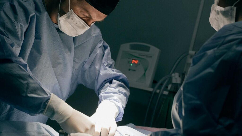
The operation process is generally performed under general anesthesia, with the patient fully asleep, due to the need for the patient’s psychological comfort, absence of pain, and the surgeon’s ability to work meticulously at a millimetric level. While the patient is in deep sleep, the surgeon clearly displays on the screen the borders where the adhesion begins, spreads, and ends under the guidance of an endoscopic camera. Different cutting and separation systems are used according to the type, thickness, and vascular structure of the area where the adhesion is located. For thin, membrane-like adhesions, cold surgical scissors that do not cause any heat damage to the tissue are preferred. The absence of heat damage is very valuable for preserving the surrounding healthy tissues.
In much thicker, harder adhesions that are highly prone to bleeding, advanced laser systems offer major advantages. Laser technology cuts the fibrotic tissue by vaporizing it with light energy, while simultaneously sealing the surrounding microscopic capillaries and stopping bleeding instantly. If the adhesion has turned into grape-cluster-like pieces of tissue called polyps inside the nose, motorized devices with a microscopic blade rotating at very high speed at the tip and a powerful vacuum are used. These devices shave and remove the faulty tissue millimetrically, leaving behind a completely flat, smooth, and healthy tissue surface. In cases where turbinate enlargement accompanies the adhesion, submucosal radiofrequency waves are used to reduce the volume of the tissues.
The technological tools used during surgery are as follows:
- Surgical scissors
- Diode laser
- Carbon dioxide laser
- Microdebrider device
- Radiofrequency probes
- Endoscopic cameras
How Is the Nasal Valve Repaired During Intranasal Adhesion Surgery?
Intranasal adhesions mostly damage the nasal valve region, which is the narrowest area of the nose, the most sensitive to lack of airflow, and the most resistant to breathing. From the perspective of the functional dynamics of the nose, the nasal valve is the keystone, the main switch of breathing. If scar tissue and adhesion have narrowed or disrupted the structure of this critical area, the problem experienced by the patient is not merely that the inside of the nose is blocked. When these patients try to take a deep breath, due to the pressure difference outside, the nasal wings collapse completely inward and close the airway like a vacuum.
The internal nasal valve is a very specific, narrow angle located between the cartilage structure forming the nasal septum and the cartilages supporting the outer side walls of the nose. Hard scar tissues that develop after adhesion reduce this angle far below what it should be. To solve this vital problem and strongly restructure the collapsed valve area, extra cartilage supports, namely grafting techniques, must definitely be used during surgery. These support cartilages to be used are usually taken from the patient’s own nasal septum, or if there is not enough material left there, from the ear or rib. These obtained cartilage pieces are placed on the nasal roof like a roof beam, mechanically widening the narrowed angle in a way that will not collapse again.
The types of cartilage supports used in valve repair are as follows:
- Spreader supports
- Butterfly grafts
- Nasal wing supports
- Umbrella grafts
- Expansion grafts
- Structural struts
Is Intranasal Adhesion Surgery Difficult After Previous Operations?
The treatment of adhesions that occur in patients who have previously undergone one or more nasal surgeries for different reasons requires much higher technical skill, experience, and attention compared to a person who will undergo nasal surgery for the first time. Due to previous unsuccessful or problematic surgeries, the natural and perfect anatomy of the nose may have been completely disrupted, tissue planes may have merged, anatomical landmarks guiding the surgeon may have been erased, and the inside of the nose may have been completely covered with dense concrete-like scar tissue. This situation resembles restoring a damaged building that is about to collapse while keeping it standing, rather than building a house from scratch.
In such challenging cases called revision, the biggest and most critical problem faced by the surgeon is that the structural material urgently needed for repair, namely healthy cartilage tissue, may have been completely used up, wasted, or melted due to infection in previous surgeries. When sufficient cartilage cannot be found to keep the nasal skeleton upright, widen the valve area, and provide support to adhesion areas opened by cutting, spare tissue sources from other parts of the body are used. These spare cartilages are shaped with fine workmanship and adapted inside the nose millimetrically.
Alternative structural materials used in revision surgeries are as follows:
- Auricular cartilage
- Rib cartilage
- Cadaver cartilage
- Synthetic grafts
- Free mucosal grafts
What Is Done After Intranasal Adhesion Surgery to Prevent the Disease from Recurring?
In intranasal adhesion surgeries, the physician performing the operation’s work certainly does not end the moment the problematic tissues are cut and the nose is opened; the truly difficult, stressful, and attention-requiring process begins exactly at this point. The most difficult part of the surgery is preventing these two fresh, bleeding, and open wound surfaces, which have been successfully separated from each other, from finding each other again and fusing during the healing phase. Scientific studies have definitively proven that performing only a rough cutting procedure during surgery and leaving the nose on its own causes the disease to recur much more severely. For this reason, immediately at the end of the operation, special pharmacological medications and physical barriers must be used simultaneously with great care in order to prevent the tissues from adhering again.
Among the strongest medical agents used for this purpose are cell-suppressing solutions with extraordinary controlling effects on wound healing. These special fluids temporarily stop the proliferation of cells called fibroblasts, which cause excessive and uncontrolled growth of scar tissue and therefore adhesions, and their production of more collagen than necessary. Immediately after the adhesion is cut and the area is cleaned, special surgical cottons soaked in this fluid are placed on the relevant area and allowed to penetrate the tissue for a few minutes. This short but effective intervention at the cellular level incredibly reduces the risk of recurrence of the adhesion.
However, medication use alone is not sufficient; building a physical wall is essential. The classic cloth tampons, kilometers long and causing great pain when removed, which were the nightmare of patients in the past, have now been completely abandoned. Instead, extremely soft, thin, and flexible silicone plates are placed inside the nose, separately into the right and left passages. These silicones create an impassable barrier between the two wound surfaces, making it physically impossible for the tissues to touch each other during the healing process. Moreover, these modern silicones have special air channels in the middle that allow breathing; thus, the patient can begin breathing comfortably through the nose as soon as waking up from surgery.
The methods and materials used to prevent recurrence are as follows:
- Mitomycin-C solution
- Silicone splints with air channels
- Bioabsorbable gels
- Hyaluronic acid coverings
- Antibiotic ointments
- Steroid drops
What Rules Should Be Followed During the Recovery Process After Intranasal Adhesion Surgery?
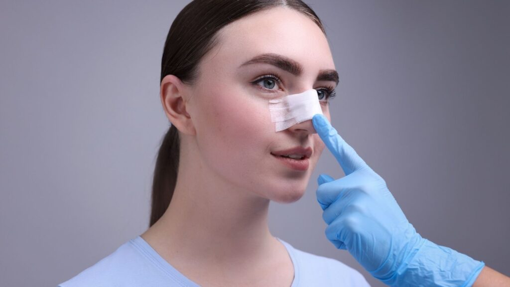
No matter how technologically perfect a surgery has been performed, the final quality and permanence of healing are directly related to how faithfully the patient follows the postoperative home care rules. The first weeks following the operation are extremely critical in order to preserve the success achieved on the operating table and prevent the adhesion from forming again. During this process, the intranasal tissues are very sensitive, edematous, and completely defenseless against external effects. The slightest mistake the patient makes in daily life may cause the wound surfaces to approach each other and months of treatment to return to the beginning.
The first forty-eight hours following surgery are the period when tissue edema and swelling are experienced most intensely. During this process, it is very important to benefit from the power of gravity. The patient using extra pillows to keep the head higher than the body while lying down lowers the blood pressure in the head region and helps the edema disperse rapidly. Since the fine capillaries inside the nose have only just been sealed, avoiding any activity that will suddenly raise blood pressure is of vital importance. Bending over to tie shoes, lifting heavy objects, straining, or blowing the nose forcefully to clear clots inside the nose causes fresh wounds to bleed and the healing process to stop. To prevent minor impacts to the nose while dressing and undressing, front-buttoned or zippered clothes should be preferred for a while instead of tight clothes that pass over the head.
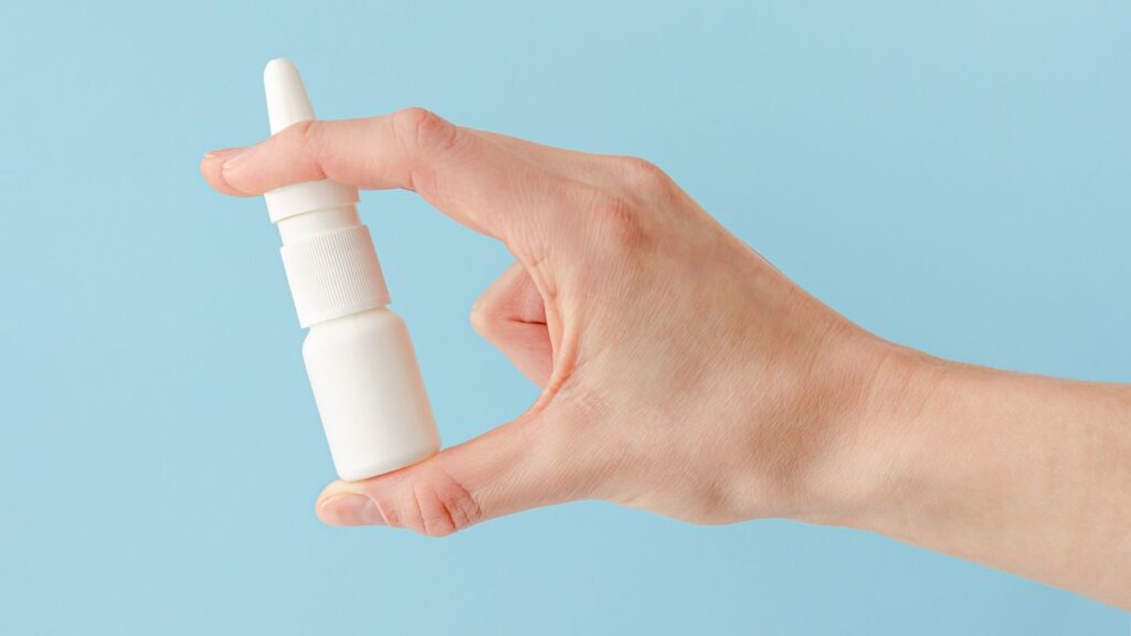
The biggest responsibility in this process is to perform intranasal cleaning without interruption. The inside of the nose should be washed several times a day under pressure with prescribed ocean waters or special irrigation kits. This procedure removes the accumulated leakage, tissue residues, and fibrin networks that would lay the groundwork for new adhesions before they dry.
Situations that must definitely be avoided during the recovery process are as follows:
- Forceful nose blowing
- Heavy sports exercises
- Excessively hot showers
- Blood-thinning medications
- Dusty environments
- Tight-collared clothes
- Sleeping face down
- Pool and seawater
What Health Problems Occur If Intranasal Adhesion Surgery Is Postponed?
Intranasal adhesions are generally perceived by patients as merely a simple nasal obstruction, a temporary loss of comfort, or a tolerable aesthetic problem. However, when this anatomical disorder is not treated in a timely and correct manner, it is the beginning of a dangerous process that deeply affects not only the nasal region but also other vital systems of the body through a chain reaction and creates serious clinical conditions. The nose being disabled means the cancellation of the body’s main air intake filter, and the cost of this condition is usually paid by essential organs such as the heart, lungs, and brain.
Among the most common and inevitable consequences encountered are resistant chronic infections. When the adhesion tissue completely blocks the millimetric natural openings of the sinuses, which are cavities inside our skull, into the nose, the mucus fluid normally produced every day inside the sinuses and cleared by swallowing cannot drain out. This closed environment filled with airless, dark, and mucus creates a perfect incubator for bacteria and fungi to multiply rapidly. Over time, these infections carry potential dangers that may progress to the eye sockets or the meninges by eroding the sinus bones from the inside. More importantly, chronic nasal obstruction forces the patient to breathe continuously through the mouth. Cold air that is not filtered, warmed, or humidified going directly to the lungs triggers lower respiratory tract diseases. Sleep apnea experienced at night drops the blood oxygen level below critical values and puts excessive stress on the heart; in the long term, this condition lays the groundwork for serious cardiovascular diseases.
The risks that may occur in case of postponement are as follows:
- Chronic sinusitis
- Sleep apnea
- High blood pressure
- Heart enlargement
- Lower respiratory tract infections
- Chronic fatigue syndrome
- Complete loss of sense of smell
- Depression and anxiety