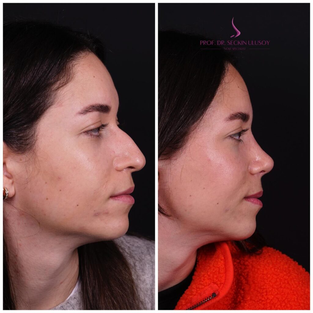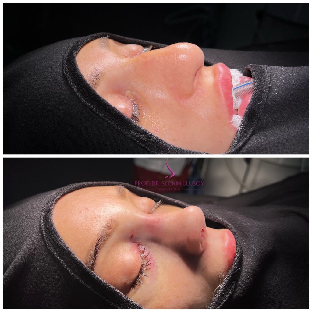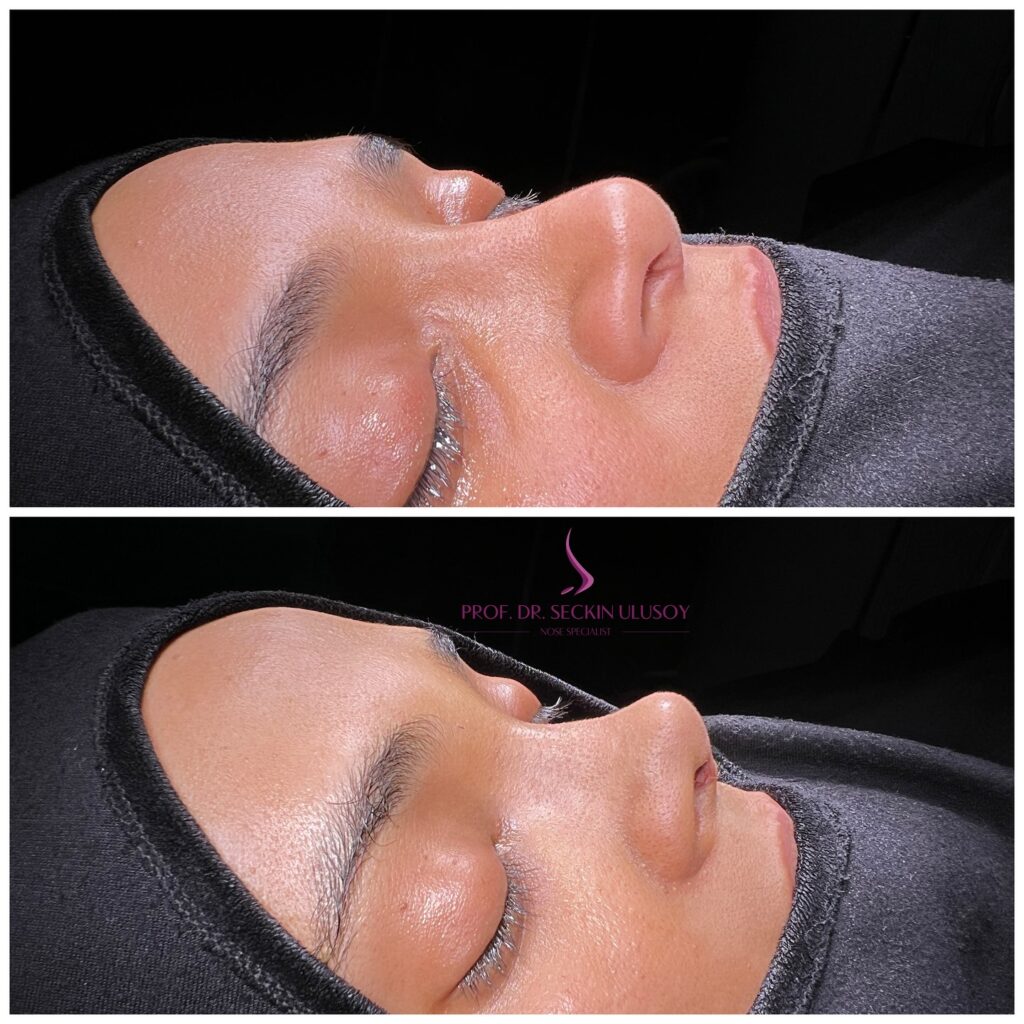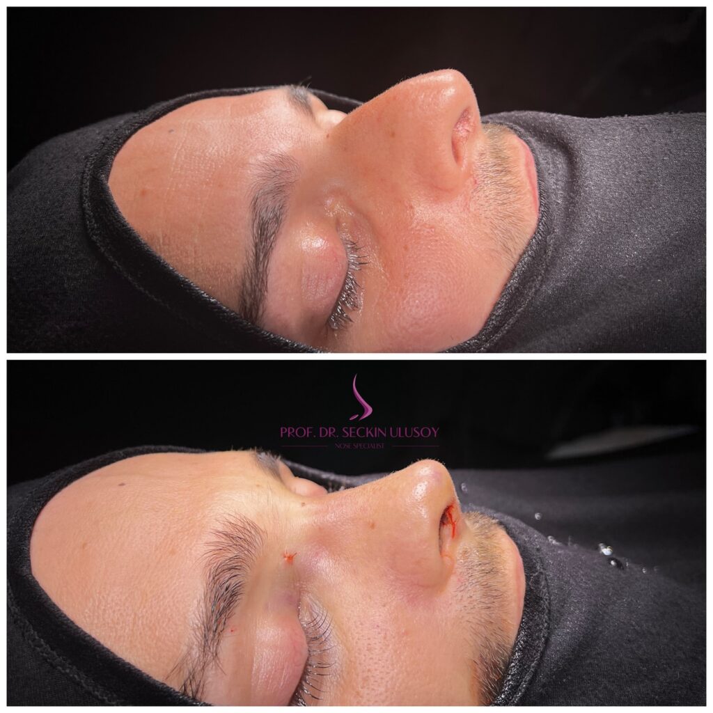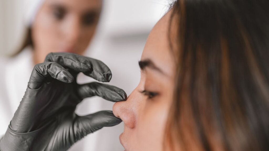
What Is Done in Nasal Obstruction Surgery?
What Is Septal Deviation and What Is Done During Its Surgery?
The septum is an anatomical structure located in the middle of the nose, consisting mostly of cartilage, with its posterior part made of bone. In order to breathe well through the nose, the septum must be straight and positioned in the midline. However, due to developmental reasons or trauma, the septum may become curved and cause nasal obstruction. The operation performed to correct this curvature of the cartilage is called septoplasty, or deviation surgery.
When deciding on surgery for deviation, the patient’s complaints and the negative effects of nasal obstruction on health and quality of life are taken into consideration as much as the examination findings of the nose.
In patients diagnosed with obstructive sleep apnea, even if there is no significant complaint of nasal obstruction, it may be necessary to eliminate the curvatures and turbinate enlargements inside the nose and provide airflow both to control apnea and to enable the use of a CPAP device.
There is no alternative other than surgical treatment in patients whose nasal obstruction complaint is determined to be caused by septal curvature (deviation).
Successful results can be achieved with standard operations performed through the nostrils and lasting approximately 20-30 minutes in curvatures that are not close to the nasal dorsum, do not involve excessive deformity in the cartilaginous part forming the anterior half of the septum, and do not include the bony framework of the nose.
In the presence of the problems described, in patients who have previously undergone nasal surgery, or in patients for whom aesthetic interventions are planned during the same operation, “open technique surgery” should be preferred.
With the surgical techniques developed today, it is possible to perform deviation and aesthetic nasal surgeries without nasal packing, which makes a significant contribution to patients’ postoperative comfort.
If patients who need surgery due to deviation are also considering aesthetic nasal surgery, the ideal approach would be to perform both operations at the same time. The ability to use excess cartilage or bone tissues that cause curvature inside the nose for various purposes in the aesthetic part of the operation is the most important advantage of performing the surgeries simultaneously.
Deviation surgeries are preferably performed under general anesthesia and last approximately 20-30 minutes with the closed technique and around 1 to 2.5 hours with the open technique, depending on the degree of the problem and whether there is a need to take cartilage tissue from the ear or rib.
Patients are generally discharged on the day of surgery or the following day.
If silicone nasal packing has been placed inside the nose, it is removed after 2-7 days. We no longer use the old cloth packs in these operations, so this is a very comfortable procedure that causes no pain to the patient. Cleansing sprays containing saline and softening drops are used for dryness and crusting that occur for 2-3 weeks until healing of the nasal mucosa is complete.
Patients must protect their nose from trauma for 6 weeks after deviation surgery. They may begin sports activities after 2 weeks. There is no harm in wearing glasses for patients who have undergone deviation surgery.

What Is Turbinate Reduction Surgery?
In the treatment of obstruction caused by enlargement of the inferior turbinates (turbinate hypertrophy), which generally causes more complaints at night, the turbinates can be reduced as much as necessary using different methods. The inferior turbinates, which play a very important role in nasal functions, should never be completely cut out and removed. Following such surgeries, which were frequently performed in the past, nasal functions are impaired, and in addition to the negative consequences on respiratory physiology, complaints such as dryness, crusting, recurrent infections, and a burning sensation inside the nose become extremely difficult and sometimes even impossible to resolve. Therefore, techniques that do not damage the functional outer mucosal surface are preferred today when reducing the inferior turbinates.
In the majority of patients with curvature of the septum to one side, enlargement occurs in the turbinates, especially the inferior turbinate, in the opposite nasal airway, which has a larger volume. Therefore, in most septal deviation surgeries, in order to obtain a satisfactory respiratory result, it is necessary to reduce the turbinates located in the nasal passage opposite the curvature with an appropriate intervention during the same operation. Otherwise, when the septum displaced to one side is brought to the midline, the previously wide side will become relatively narrower, and the turbinates that had adapted to the preoperative volume of this part will cause obstruction. You can contact us to get information about nasal obstruction surgery.
The most commonly applied methods for reducing the inferior turbinates are reducing the volume of submucosal tissue without damaging the mucosa using radiofrequency and, in cases where the bony part of the turbinates is also enlarged, reducing the soft tissue and bone tissue beneath the mucosa by shaving them (endoscopic turbinate reduction).
An air cell may be present within the middle turbinate as a structural variation in approximately 20% of the population (concha bullosa). Enlargement of this air cell during development prevents airflow in the middle nasal passage and may also cause chronic sinusitis due to obstruction of the sinus drainage channels located on the outer wall of the passage. Appropriate endoscopic surgical methods are also frequently applied to middle turbinates that cause such problems. Although complete removal of the middle turbinates, which are not as critical as the inferior turbinates in terms of breathing comfort, does not cause a serious problem in nasal physiology, it is generally preferred not to remove these structures completely because they serve as anatomical guides for possible future sinus interventions.
Reduction of the Inferior Turbinate with Radiofrequency:
It is based on the principle that, after radiofrequency energy is delivered with a special device into the soft tissues rich in blood vessels beneath the inferior turbinate mucosa, the heat damage formed in the tissues heals through contraction and hardening. Following this procedure, hard crust tissue may form for 15 days due to the swelling that develops or the tissue reaction on the mucosal surface, so the patient will achieve the actual improvement in breathing performance after 6 weeks. In patients who do not require intervention for another pathology, inferior turbinate radiofrequency treatment can also be performed under local anesthesia in office conditions.
The turbinate radiofrequency procedure, which takes approximately 5-10 minutes, can also frequently be performed during nasal surgeries carried out for aesthetic or functional purposes, surgeries for chronic sinusitis, and surgeries performed for another reason in patients with nasal obstruction complaints.
Since no incision is made in the turbinate during this procedure, there is no postoperative bleeding, and therefore nasal packing is not required. Following good local anesthesia, patients can immediately return to their daily activities after this procedure, during and after which no pain is felt.
In patients who achieve successful results with radiofrequency, the inferior turbinates may enlarge again within 6 months to 1 year if conditions such as allergic rhinitis or vasomotor rhinitis are present, if patients are continuously exposed to dry and polluted air, cigarette smoke, or cold environments, or if they use vasodilator medications. In such cases, the procedure may rarely need to be repeated.
The turbinate radiofrequency procedure is one of the procedures most frequently preferred by patients and physicians due to advantages such as its high success rate, not requiring nasal packing, being performable under local anesthesia, and not causing bleeding.
Endoscopic Inferior Turbinate Reduction:
It is the procedure of partially removing the soft tissues beneath the inferior turbinate mucosa and the turbinate bone. This method is generally applied in cases where the bone within the turbinate is an important factor in turbinate enlargement, where the desired result cannot be achieved with radiofrequency applications, where the turbinate is excessively large or deformed, or where radiofrequency treatment is not preferred because of the possibility of recurrence.
The procedure is performed under general anesthesia and takes approximately 5-10 minutes when performed with microdebrider technology. Since the newly developed microdebrider devices for the turbinates are very small, the integrity of the mucosa is not disrupted during the procedure, and therefore endoscopic reduction surgeries can now also be performed without nasal packing.
Following endoscopic turbinate reduction surgery, which is a highly effective method, significant reduction occurs in the turbinates. With the help of new microdebriders, the posterior parts of the inferior turbinates can also be reached, and successful reduction can be performed in inferior turbinates whose posterior parts are enlarged or deformed. In some patients in this condition, partial removal or radiofrequency application to the posterior part of the inferior turbinate may also be preferred.


Why Is the Nasal Valve Important in Nasal Obstruction and How Is Valve Surgery Performed?
Nasal valve insufficiency is observed in two different types: internal or external.
Internal insufficiencies result from narrowing of the airflow passage in the valve region due to curvatures close to the nasal dorsum in the anterior part of the cartilaginous septum. In treatment, in addition to exposing this region with closed-technique deviation surgery and correcting the curvature, the valve region may be widened with the help of cartilage grafts when necessary to facilitate airflow.
The main problem in external valve insufficiency is that the nasal wings collapse inward during breathing and narrow the airflow passage. This problem results from the cartilage structures within the nasal wings being structurally curved in the opposite direction or becoming weakened due to aesthetic surgeries, trauma, aging, and similar causes, making them unable to withstand the vacuum effect created by airflow. In treatment, techniques such as supporting the weak areas with cartilage pieces, correcting the cartilage angle that causes narrowing, and preventing collapse with the help of sutures are used.